© Benaki Phytopathological Institute
Plant parasitic nematode fauna in citrus orchards in Iran
103
Male
: Not found.
REMARKS
According to the morphological charac-
ters and morphometric data given in Ger-
aert (2013), there were no differences be-
tween the Iranian population of
P. musii
and
the original description. However, the post-
uterine sac length is shorter (
14-18.5
vs
22-
30 μm).
The species was originally
recovered
from the rhizosphere of banana
and de-
scribed by
Choudhury and Phukan,
1989
from Assam, India (
Geraert, 2013)
. In a study
of nematode community associated with
banana in Assam, India, Deori
et al.,
2014
found that
P. musii
was one of the predom-
inant nematode species around the banana
rhizosphere.
In the present study, this species was re-
covered from 9.8% of soil samples from the
rhizosphere of orange and tangerine in the
vicinity of Shush city, Khuzestan province,
Southwestern Iran. This is the first record of
P. musii
for the nematode fauna in Iran.
Psilenchus hilarulus de Man (1921)
Figure 5
MEASUREMENTS (Table 3)
Iranian population of
P. hilarulus
is in
morphological and morphometric agree-
ment with the original description (Geraert,
2008). However, the length of female body is
shorter (640-845
vs
890-1150 μm).
This spe-
cies
has been reported from the citrus rhizo-
sphere in Mazandaran province, Iran (Divsa-
lar
et al.,
2011). Also, has been reported from
the rhizosphere of sugarcane in Khuzestan
province, Iran (Kheiri, 1995). In the pres-
ent study, this species was recovered from
20.5% of soil samples from the rhizosphere
of orange, lemon and sour orange in the vi-
cinity of Shush, Dezful, Ramhormoz, Bagh-
malek and Ramin cities, Khuzestan province,
Southwestern Iran.
Tylenchulus semipenetrans Cobb (1913)
Figure 6
MEASUREMENTS (Table 4)
Characters measured in Khuzestan pop-
ulation of
T.
semipenetrans
are consistent
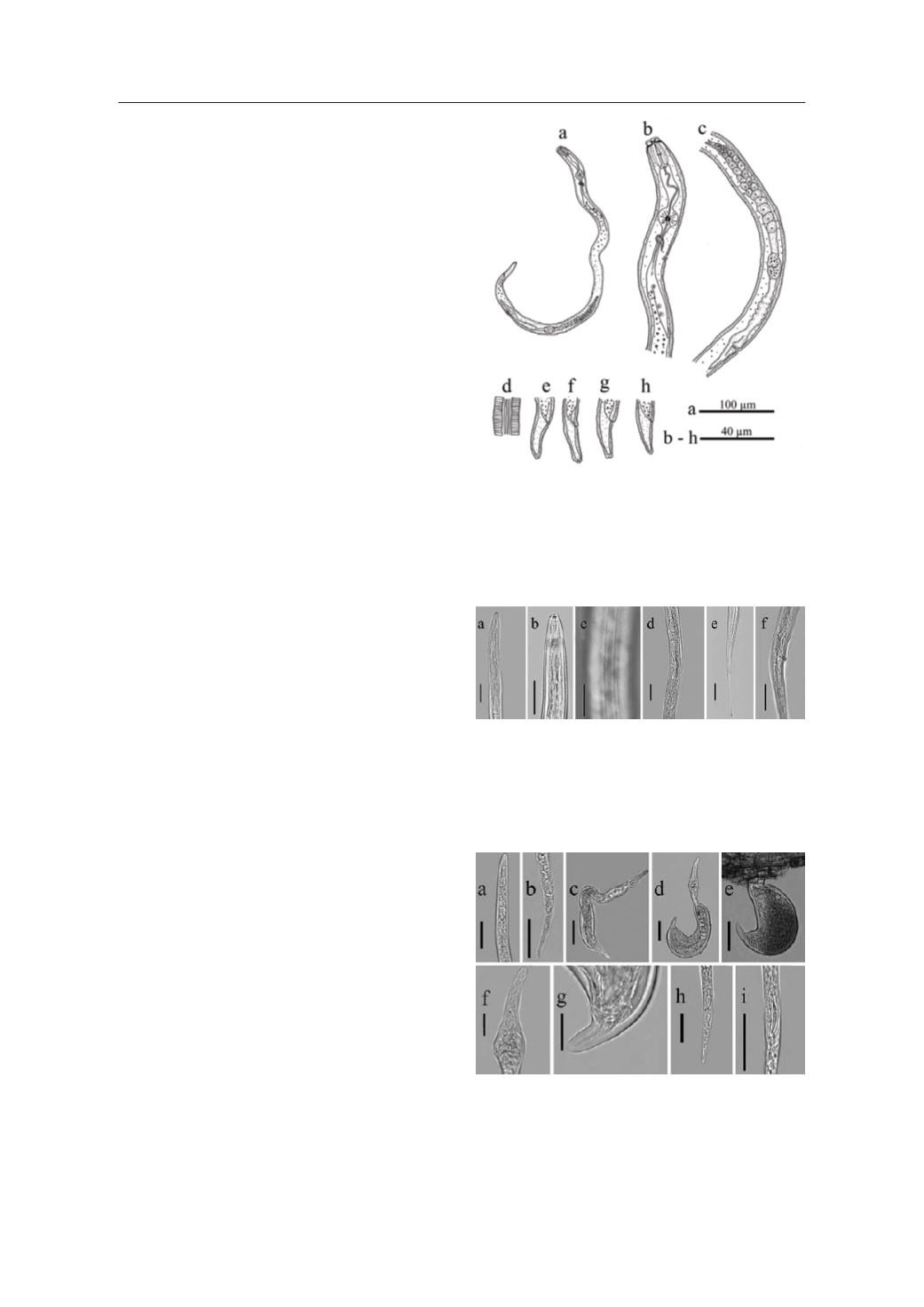
Figure 4.
Female of
Pratylenchus musii
. a: Entire body; b: An-
terior region; c: Vulval region; d: Lateral field at mid-body; e-h:
Tail.
Figure 5.
Psilenchus hilarulus.
a, b: Anterior region; c: Lateral
field at mid-body; d: Vulval region; e: Female tail; f: Male tail.
(Scale bars: 20 μm).
Figure 6.
Tylenchulus semipenetrans.
a: Anterior region of Ju-
venile; b: Posterior region of Juvenile; c: Immature female; d,
e: Mature female; f: Anterior region of female; g: Posterior re-
gion of female; h, i: Male tail. (Scale bars: a, b = 20 μm, c-e =
50 μm, f-i = 20 μm).


